


New Approach to Osteoarthritis Treatment
Researchers at the Yale School of Medicine have discovered that lacosamide, an existing U.S. Food and Drug Administration-approved epilepsy medication, can be repurposed to target osteoarthritis by easing pain while helping reverse physical cartilage damage in preclinical studies, as reported by ScienceAlert. Osteoarthritis occurs when the cushioning and lubricating cartilage between bones wears away over time. Current standard medical treatments can help manage associated symptoms, but there is currently no approved pharmacological fix to reverse structural joint deterioration, leaving joint replacement surgery as the primary advanced option for severe progression.
The findings, published in the journal Bioactive Materials, demonstrate that testing in mouse models and human cartilage cells in laboratory settings showed the drug is capable of encouraging cartilage repair, slowing deterioration, and reducing pain simultaneously. Because lacosamide has already cleared human safety evaluations for epilepsy, its established regulatory profile could potentially shorten the timeline required for clinical trials, offering fresh hope for hundreds of millions of people worldwide affected by the condition.
The Role of the Nav1.7 Protein Target
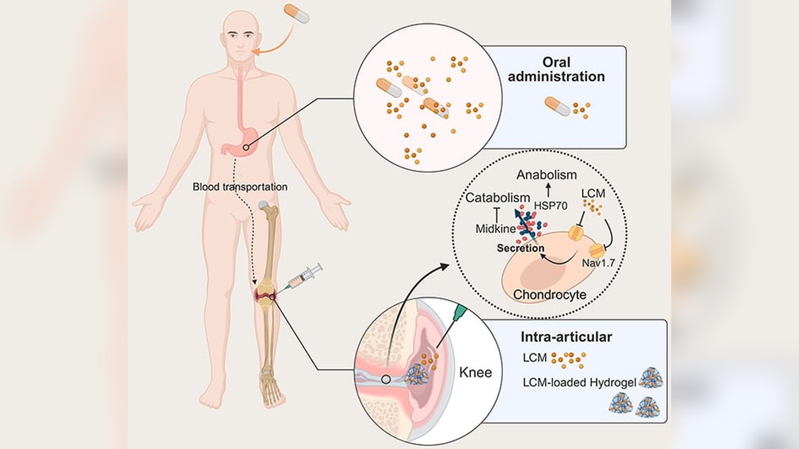
The research team was led to lacosamide because it functions as a sodium channel blocker. In prior work, scientists identified a specific sodium channel and protein known as Nav1.7 that is involved in transmitting pain signals to the brain while also regulating how joint cartilage is built up and broken down. Nav1.7 is active inside chondrocytes, which are specialized cells that manage the balance of removing old tissue and synthesizing new material.
In joints affected by osteoarthritis, Nav1.7 activity increases beyond normal levels, disrupting this balance and tipping the scale toward cartilage loss. “When Nav1.7 becomes dysregulated, it contributes to both joint degeneration and pain,” explains musculoskeletal biologist Chuan-Ju Liu, who serves as the study’s principal investigator and Charles W. Ohse Professor of Orthopaedics & Rehabilitation at Yale. By blocking this dual-acting protein target with lacosamide, the researchers aim to quiet pain-sensing nerves while signaling chondrocytes to stop breaking down tissue and start repairing it.
Optimizing Dosage and Hydrogel Delivery
During laboratory evaluations, the team discovered that lacosamide’s therapeutic benefits depended heavily on dosage concentration. Rather than following a more-is-better trajectory, the drug required a specific optimal concentration range to encourage cells to produce cartilage-building proteins while suppressing destructive processes. “This tells us the system is finely tuned,” Liu notes, emphasizing that the ideal low concentration restored balance without overcorrecting.
Furthermore, direct injection into affected joints proved more effective than oral administration, but pure liquid treatments tended to dissipate quickly from the joint cavity. To solve this challenge, the researchers packed lacosamide inside a specialized hydrogel. According to the Yale School of Medicine, this hydrogel functions as a local reservoir that holds the medication in place and releases it slowly over a period of a month or longer.
Broader Cellular Signaling Mechanisms
Beyond blocking Nav1.7 directly, further investigation revealed that lacosamide changes cellular communication pathways. The treatment stimulated the release of two beneficial signaling proteins, HSP70 and midkine. HSP70 helps cells respond to environmental stress and supports tissue repair, while midkine assists in regulating inflammation and protecting joint tissue from further degeneration.
These secondary proteins help create a supportive biochemical environment that extends beyond individual chondrocytes to influence the entire joint tissue matrix. While these preclinical outcomes in cellular models and mice represent a major scientific step forward, the therapy still requires rigorous clinical testing in human patients before it can be considered for public medical use.